The measurement of pupil diameter is an important component of neuro–examinations since it helps in assessing the status of a patient’s neurological health. Years later, there have been improvements in technology that have seen changes in how pupil size, velocity of the pupils, and other related activities are measured. This blog discusses these innovations, specifically the tools and technologies that have made a major difference in neurological examinations.
Introduction to Pupilometers
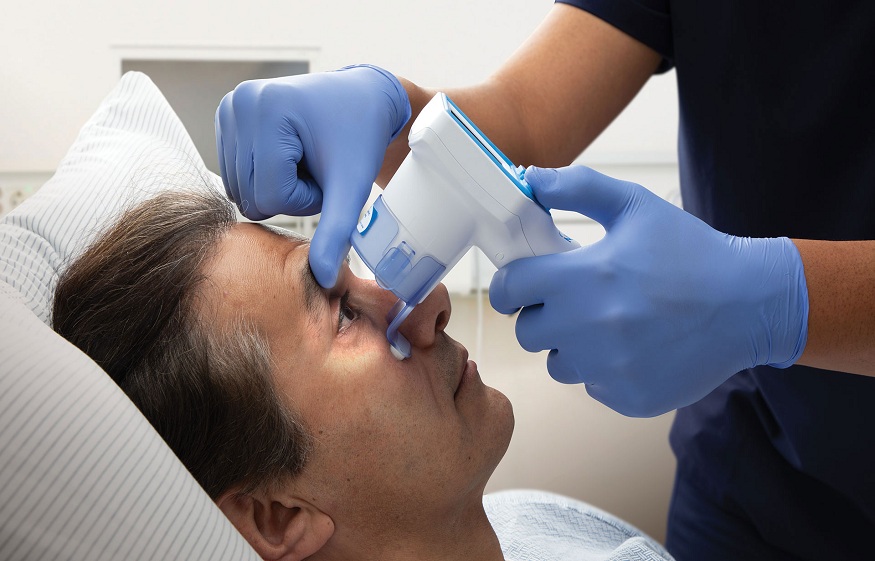
In this regard, the innovation of the pupilometer is one of the most impactful steps made in measuring the pupil diameter. A pupilometer is an electronic device that is meant to assess the size and responsiveness of the pupils with close precision. These devices offer several advantages over traditional methods, including These devices offer several advantages over traditional methods, including:
Accuracy: Pupilometers are accurate in assessing pupil size compared to visual estimation and are less likely to have measurement errors.
Consistency: The use of electronics in the measurement of the examinations helps in maintaining standards in the results of various examinations and examiners.
Ease of Use: Pupilometers are also easy to operate and thus healthcare professionals can conduct the assessments with ease.
New Developments in Measuring Pupil Dilation Velocity
Pupil diameter velocity, the rate by which the pupil of the eye changes its size in response to light, is another significant aspect considered in neuro exam. Such deviations can also point towards neurological disorders like issues with the dilation velocity of the pupils. This parameter can be measured with great accuracy and relative ease, and recent innovations have made it even better.
Dynamic Pupillometry
Static pupillography is a method to measure pupil size in response to light stimuli, this is done in a real-time manner. New-generation dynamic pupillometers utilize a high-speed camera and complex software to monitor changes in the size and shape of the pupils. It is useful in measuring Pupil Dilation velocity making it a good tool in neurological tests and diagnosis.
Video Pupillometry
The innovation of video pupillometry is another significant step forward in determining the size of the pupils and their response patterns. In this method pupil response to light is captured on the video camera and later reviewed on the computer with the help of certain software. Video pupillometry offers several benefits, including Video pupillometry offers several benefits, including:
Comprehensive Analysis: This makes the video recording useful since one gets a good view of the pupil’s behavior pattern when being taught.
Non-Invasive: It is painless and less intrusive to patients, hence suitable for repeated measurements on the same patient.
Enhanced Diagnostic Capabilities: Because video pupillometry can track individual pupillary responses, it may be able to identify changes in pupil behavior that other approaches do not.
Integration of Neurological Tools
More advanced neurological tools have been integrated into the process to improve the degree of accuracy with which pupil diameter is measured. One of the tools is the Neurological Pupil Index (NPi).
Neurological Pupil Index (NPi)
The NPi is an index score computed with data obtained from pupillometry, which can standardize pupil reactivity measures. This index is arrived at based upon the application of various algorithms that take into account factors such as the size of the pupils, the velocity of their dilation, and the latency of their constriction respectively. The NPi offers several advantages:
Standardization: The NPi offers the means to measure the condition more uniformly, minimizing the variation between different examiners and increasing the validity of the evaluations.
Early Detection: While the existence of the NPi can identify several parameters at the same time, it can detect signs of early neurological decline that are not revealed through other manual evaluations.
Monitoring: The NPi is beneficial in tracking a patient’s neurological state alteration within a specific period, and thus identifying if the patient’s condition worsens or improves.
Conclusion
Technological developments in the measurements of the pupil diameter have greatly progressed the neuro exam. Pupilometers or tools that measure pupil size and dynamic and video pupillometry with the inclusion of the NPI as brought significant changes to how healthcare workers assess and interpret the size and reactivity of pupils. Based on these findings and due to the ever-advancing technology, there is hope for further development of neurological assessments that are more accurate, reliable, and easily accessible to enhance patient care.

